Monday 30 January 2017

What is a Horned Lizard?

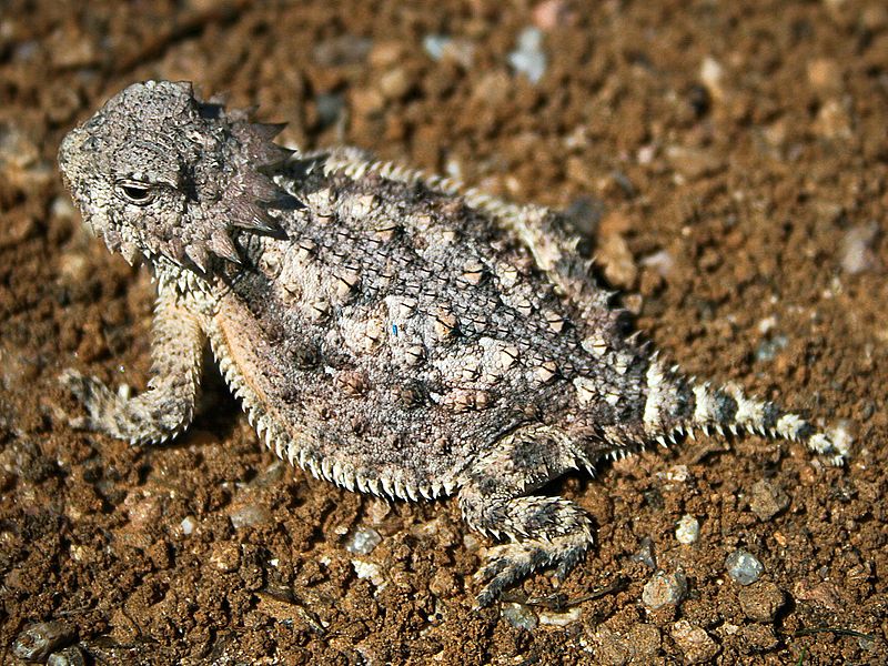
Horned lizard 032507 kdh.jpg

Horned lizards are a genus (Phrynosoma) of lizards which are the type genus of the family Phrynosomatidae. The horned lizard has been affectionately called a "horny toad", or "horned frog", though they are not moist-skinned toads or frogs. The common names come from the lizard's flattened, rounded body and blunt snout, which make it resemble a toad or frog (Phrynosoma means "toad-bodied"), as well as its tendency, in common with larger true frogs and toads, to move sluggishly, making them easy to hand-catch (such slow, undramatic movements may also avoid triggering attacks by predators, discussed later in this article). They are totally adapted to desert areas. The spines on its back and sides are made from modified reptile scales which prevent the water loss through the skin, whereas the horns on the heads are true horns (i.e. they have a bony core). Of 15 species of horned lizards in North America, eight are native to the United States. The largest-bodied and most widely distributed of the US species is the Texas horned lizard.


        Horned lizards use a wide variety of means to avoid predation. Their coloration generally serves as camouflage. When threatened, their first defense is to remain still to avoid detection. If approached too closely, they generally run in short bursts and stop abruptly to confuse the predator's visual acuity. If this fails, they puff up their bodies to cause them to appear more horned and larger, so that they are more difficult to swallow. At least eight species (P. asioP. cornutumP. coronatumP. ditmarsiP. hernandesiP. orbiculareP. solare, and P. taurus) are also able to squirt an aimed stream of blood from the corners of the eyes for a distance of up to 5 feet (1.5 m).[1][2][3][4] They do this by restricting the blood flow leaving the head, thereby increasing blood pressure and rupturing tiny vessels around the eyelids. This not only confuses predators, but also the blood tastes foul to canine and feline predators. It appears to have no effect against predatory birds. Only three closely related species (P. mcalliiP. modestum, and P. platyrhinos) are certainly known to be unable to squirt blood.[2] To avoid being picked up by the head or neck, a horned lizard ducks or elevates its head and orients its cranial horns straight up, or back. If a predator tries to take it by the body, the lizard drives that side of its body down into the ground so the predator cannot easily get its lower jaw underneath.
                       Image result for horned lizard

Horned lizards protect themselves in many ways, including breaking the blood vessels in their eyes and shooting blood up to 4 feet in the air to distract predators, according to the Missouri Department of Conservation. They also eject small amounts of blood from the inner corners of their eyes to confuse predators.


Can turtles breathe through their butts?

Turtles Breathe Through Their Butts

flicker Brent Myers
Red Eared Slider (Photo: Brent Myers, Flicker Sharing)

Vent Breathing Through Cloacal Bursae

In a previous post I talked about the difference between hibernation and brumation in turtles. Essentially turtles don't sleep all winter, they have punctuated periods of activity. However, turtles do not brumate under water,  they usually dig burrows or bury themselves in leaf litter or mud to overwinter. One of the questions I'm asked frequently is,  "How do turtles breathe while they are buried?" A similar question was, "How can that turtle breathe while napping under water?" (This was usually aimed at a local terrapin that lived in a tank in the education center). The answers are similar.

Turtles can respire, or breathe, in three main ways:

1. Through their lungs. Turtles are vertebrates and they have lungs, which is their primary means of respiration. This is why sea turtles can drown if they don't come up for air. The crazy thing is, their shell is their rib-cage. Their lungs, guts, organs, legs, everything sit inside their rib cage. Look carefully at the inside of a an empty turtle shell and you can see the fused rib sutures.
4382043091_89b20c3f83_o
Inside an aquatic turtle shell (Photo: Flicker Sharing, msmecomber).
The challenge of this type of outer-rib-cage-shell is that unlike you and I, they can't take a deep breath in, expanding their body cavity and lungs, to take a breath like humans do.
I was once asked if a turtle moving its throat was a way of breathing or pumping air into its lungs. You can find LOTS of misinformation about this on the internet. Research has shown that bucal (cheek) pumping and gular (throat) pumping does not correlate to lung inflation (check out this great paper from U Mass Amherst to learn more and see awesome X-rays). Throat movement, in turtles, is more likely associated with smelling or olfaction. Scientists were partly cued into the "smelling" idea because turtles still pump their throats under water, even when they can't take oxygen into their lungs (they plug their throats with their tongue). Throat pumping under water allows  for aquatic smells  to enter through the nose and mouth even though their throat is closed for diving. This leads to point #2 below.
2. Through their mouths and throats. Stick with me here, and don't get confused. I mentioned earlier that turtles pump their throats and mouths, but it's not pumping air into their lungs. However,  some species of aquatic turtles can absorb oxygen through their mouth/throat system. If an aquatic turtle is under water it can move its throat and mouth to take in water to smell and also absorb oxygen across the surfaces inside its mouth/throat complex. The same is true above water. The membranes of the throat and mouth are usually moist, which also allows for oxygen diffusion.
Eastern_Box_Turtle_8680
The cloaca of a turtle is the "common poop chute" where poo, pee, eggs, and mating all occur. This is a female box turtle laying eggs (Photo: Wiki commons).
3. Through their butts. Yes, some species of aquatic turtles can breathe through their butts. Now this isn't like the butt-snorkel idea that I wrote about with how mosquito larvae breathe (tubes in their butts held at water level). This much more about oxygen absorption across tissues, and less like breathing through a snorkel or nose. Turtles have what I call the "common poop chute," meaning they poop, pee, mate, and lay eggs through one hole, technically called a cloaca (clo-ay-ka) or vent. In some species of aquatic turtle there are structures called cloacal bursae, which are essentially highly vascularized (filled with blood vessels) tissues which allow oxygen to be absorbed across them. In some species of turtles there are many ridges or folds lining the vents, which also allow for oxygen absorption. How much can a turtle breathe through it's butt?  Well, this varies, usually not a lot (>20%). Billy's little turtle in the aquarium isn't going to be OK if you don't let it come up for air, it needs to breathe through its lungs. Of course there are always exceptions, mostly with soft shelled turtles.
E_albagula_4
Australian white-throated snapping turtle (Photo: Wiki Commons).
The Australian White-throated snapping turtle, also called the "bum-breathing turtle" can get nearly 70% of its oxygen through its cloacal bursae, or butt.   Unfortunately this species is critically endangered and may not be around long.
No matter how you shake it, turtles are amazing survivors. Their natural adaptations for respiration are fascinating. If you're leading a lesson or unit on turtles, consider integrating information about turtle respiration. It's easy enough to throw a turtle shell into a trail-pack. You can use it to create a "filler" activity while waiting for groups, or stragglers on a hike, by asking students to compare how they breathe in and out (filling their lungs) to how turtles breathe. It's up to you to add the butt breathing or not, but the kids will LOVE it!
1093

Butterflies taste with their feets??

Butterflies taste with their feet

Butterflies taste with their feet as their taste sensors are located there. They can taste it by just standing on their food. They don't have mouths that allow them to bite or chew, instead they have a long straw-like structure called a proboscis which they use to drink nectar and juices. But the proboscis doesn't have any taste sensors. A butterfly's taste sensors are located on the bottom of its feet. When not in use, the proboscis remains like a garden hose. By standing on a leaf, the butterfly can taste it to see if it the leaf they sit on is good to lay eggs on to be their caterpillars’ food or not.


In the photo above, you can see the proboscis of the recently emerged monarch butterfly. Notice that it is in two pieces and has a forked appearance. As soon as it emerges, the butterfly begins working on the proboscis with two palpi (found on each side of the proboscis), forming it into one tube. It must do this successfully in order to be able to nectar.
Butterflies fly between 5 and 30 miles an hour. People who study butterflies are called Lepidopterists. A butterfly can lay up to 500 eggs. Butterflies flap their wings quite slowly, usually from 5-20 beats per second. Butterflies are important pollinators. They come fourth after beetles, flies, and bees. Some butterflies have wingspans of 11 inches.
Butterflies cannot fly if their body temperature is less than 86 degrees

As a caterpillar, they will increase up to several thousand times in size before pupating (process in which caterpillars transform into adult butterflies in a structure called a chrysalis )
                                                         Related image

What mammal that can fly?

                           BATS
Related image


     Bats are the only mammals that can achieve true flight. Their wings are thinner and more delicate than those of birds. There are some other mammals that can glide through the air, but this is not considered flying.
     There are more than 900 species of bats throughout the world, though some estimates range up to 1,200. Bats range in size from very small, weighing just a few grams, to the largest recorded size, weighing up to 2.2 pounds. The flying fox is the largest of the flying mammals and weighs up to 2.2 pounds, with a wingspan of up to 6 feet. Bats are a varied species, and though the species are mostly nocturnal, they have been rarely known to be active during the daytime. Although bats have notoriously bad eyesight, they use echolocation and are graceful, accurate fliers even in the dark of night or caves. The eating habits of bats range from blood and insects to fruits.

Related image

Gliding animals are relatively common, but are limited to small mammals, such as squirrels and small possums. Gliding mammals do not have wings, but rely on extra skin between their limbs to catch the air. These mammals leap from high places and ride the air to the next landing area. They typically only glide short distances, such as between two trees. Human BASE jumpers base their glide suits off of this type of mammal.

What fruit has its on the outside??

                                                         STRAWBERRY
Related image
      Strawberries are the only fruit that has seeds on the outside. The fruits are high in vitamin C and are a significant source of folic acid, which is essential for fetal development.

        While it's true that strawberries are covered in tiny yellow seeds, the red edible part of the strawberry is not actually a fruit. The dried seeds on the outside of the strawberry are the actual fruit.The fleshy part of the strawberry is a part of the flower located under the ovary, known as the receptacle or accessory fruit. This part of the flower swells and turns bright red to attract animals to eat and distribute the seeds. These small seed-like dried fruits are known as achenes.

Strawberry in fields. Mahabaleshwar. India - Dinodia/age fotostock/Getty Images

Strawberries: Contrary to popular belief, the sweet red flesh of the strawberry is not the fruit. It is a part of the flower called the receptacle that is located below the ovary. The receptacle swells to attract animals so that they will eat and scatter the true fruit. These are the small yellow seed-like parts, which is a dry fruit called an achene.The seed you see in strawberry is an achene.It's called an Accessory fruit and strawberries are not the only ones, also they aren't proper seeds either, they are called Achene.

Do penguins have knees??

                                                    PENGUINS
                     
        A penguin’s leg is composed of a short femur, knee, tibia and fibula. The upper leg bones are not visible as they are covered in feathers giving penguins a very short legged appearance. 


     Watching a penguin waddling around, it would be easy to assume they can’t bend their legs, but they can. A penguin’s leg has a short femur, knee, tibia, and fibula. The legs just look short and stubby because the upper leg is hidden by feathers.
    Penguins do have knees, but they are covered in feathers, so they are not visible. A penguin's leg has four parts: the short femur, knee, tibia and fibula.
    Because their upper leg bones are covered, penguins appear to have very short legs. Their waddling walk also makes it seem they are unable to bend their legs. Actually, some penguins can run quite fast, such as the Gentoo penguin which has been clocked at a top speed of 22 mph. Since penguins spend almost 75 percent of their lifetime in the water; their knees help them swim. Penguins live almost exclusively in the Southern Hemisphere. The Galapagos penguin, which lives near the equator, is the only species that ventures into the Northern Hemisphere.

                Do penguins lay eggs?                    

     Though you might not be able to tell from their plump bellies and adorable waddles, penguins do, in fact, have knees! They have the same leg bones as most other bipedal vertebrates, but certain adaptations have made walking a bit harder for them.
Scientists have determined that walking costs much more energy for penguins compared ot their body weight than it does for other land animals, and have concluded that this is a result of various adaptations that increase their mobility under water. Indeed, penguins are exceptionally fast and maneuverable swimmers, and all of their prey (and predators) live underwater. Their knees are usually hidden by their feathers.

Are kangaroos have 3 vaginas??

Kangaroos Have Three Vaginas, So Here Is How That Works


marsupials, including the most popular one, the kangaroo, are equipped with a total of three vaginas. Three vaginas, two uteri, all there to serve their own joey-bearing purpose. For one thing, can you imagine their periods, you guys? Crazy! For another, imagine how hilariously scared all those anti-choice lawmakers would be when they got a load of this information if it applied to human vaginas! We’d probably have to stop being women altogether! Because it’s just too many vaginas for people to understand!
The triple marsupial vagina was discussed on the British documentary Inside Nature’s Giants. On that show, anatomists conduct dissections of some of nature’s larger beasts to see how their guts work. In this case, they checked out the ladyguts of kangaroos, for the sheer heck of it. Or because they read something written on the male kangaroos’ bathroom wall and became curious.
What they found was a decidedly more complicated reproductive system than appears in other mammals. It looked something like this:
I’ll bet you’d been hoping for a chart! That’s a screenshot from the documentary, explaining the functions of a marsupial’s three vaginas. The side-vadges are where sperm — from the males’ two-pronged penises — travel up, the middle-vadge is where the baby joey slides down to develop externally in the mother’s pouch after growing to the size of a jellybean in one of her two uteri. Fun fact: Female kangaroos can be “perpetually pregnant” because of how their plumbing works. Because once one joey has left a uterus for the pouch, another embryo can go ahead and start developing internally. So, basically, kangaroos can be pregnant forever. To say nothing of the joeys who have already left the pouch and need to be cared for. Let’s all be glad we’re not kangaroos.
However, let’s be slightly jealous, because the anatomy of a marsupial doesn’t allow for a large fetus to pass through it, and that’s why the joeys are so tiny when they’re “born” (about half an inch long for kangaroos) and start climbing to the pouch, where they grab on to nurse for the next few months of development. No pushing for these mommies.

What is an Octopus??

                                                            OCTOPUS
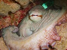
Octopus2.jpg
             Octopuses are characterized by their eight arms, usually bearing suction cups. The arms of octopuses are often distinguished from the pair of feeding tentacles found in squid and cuttlefish.[18] Both types of limb are muscular hydrostats.
              Octopuses can be divided into two suborders, the Incirrina (or Incirrata) and the Cirrina (or Cirrata). The incirrate octopuses are distinguished from the cirrate octopuses by their absence of "cirri" filaments (found with the suckers), as well as by the lack of paired swimming fins on the head. Unlike most other cephalopods, the majority of octopuses – those in the Incirrina – have almost entirely soft bodies with no internal skeleton. They have neither a protective outer shell like the nautilus, nor any vestige of an internal shell or bones, like cuttlefish or squid. The beak, similar in shape to a parrot's beak, and made of chitin, is the only hard part of their bodies. This enables them to squeeze through very narrow slits between underwater rocks, which is very helpful when they are fleeing from moray eels or other predatory fish. The octopuses in the less-familiar Cirrina suborder have two fins and an internal shell, generally reducing their ability to squeeze into small spaces. These cirrate species are often free-swimming and live in deep-water habitats, while incirrate octopus species are found in reefs and other shallower seafloor habitats.
Octopuses have a relatively short life expectancy, with some species living for as little as six months. Larger species, such as the giant pacific octopus, may live for up to five years under suitable circumstances. However, reproduction is a cause of death: males can live for only a few months after mating, and females die shortly after their eggs hatch. They neglect to eat during the (roughly) one-month period spent taking care of their unhatched eggs, eventually dying of starvation. In a scientific experiment, the removal of both optic glands after spawning was found to result in the cessation of broodiness, the resumption of feeding, increased growth, and greatly extended lifespans.[19]
Grimpoteuthis discoveryi, a finned octopus of the suborder Cirrina
Octopuses have three hearts. Two branchial hearts pump blood through each of the two gills, while the third is a systemic heart that pumps blood through the body. Octopus blood contains the copper-rich protein hemocyanin for transporting oxygen. Although less efficient under normal conditions than the iron-rich hemoglobin of vertebrates, in cold conditions with low oxygen pressure, hemocyanin oxygen transportation is more efficient than hemoglobin oxygen transportation. The hemocyanin is dissolved in the plasma instead of being carried within red blood cells, and gives the blood a bluish color. The octopus draws water into its mantle cavity, where it passes through its gills. As molluscs, octopuses have gills that are finely divided and vascularized outgrowths of either the outer or the inner body surface.

Defense

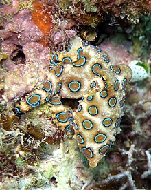
Greater blue-ringed octopus (Hapalochlaena lunulata)
The octopus's primary defense is to hide or to disguise itself through camouflage and mimicry.[39] Octopuses have several secondary defenses (defenses they use once they have been seen by a predator). The most common secondary defense is fast escape. Other defenses include distraction with the use of ink sacs and autotomising limbs.
Most octopuses can eject a thick, blackish ink in a large cloud to aid in escaping from predators. The main coloring agent of the ink is melanin, which is the same chemical that gives humans their hair and skin color. This ink cloud is thought to reduce the efficiency of olfactory organs, which would aid evasion from predators that employ smell for hunting, such as sharks. Ink clouds of some species might serve as pseudomorphs, or decoys that the predator attacks instead.[40]
The octopus's camouflage is aided by certain specialized skin cells which can change the apparent color, opacity, and reflectivity of the epidermis. Chromatophores contain yellow, orange, red, brown, or black pigments; most species have three of these colors, while some have two or four. Other color-changing cells are reflective iridophores, and leucophores (white).[41] This color-changing ability can also be used to communicate with or warn other octopuses. The highly venomous blue-ringed octopus becomes bright yellow with blue rings when it is provoked. Octopuses can use muscles in the skin to change the texture of their mantle to achieve a greater camouflage. In some species, the mantle can take on the spiky appearance of seaweed, or the scraggly, bumpy texture of a rock, among other disguises. However, in some species, skin anatomy is limited to relatively patternless shades of one color, and limited skin texture. It is thought that octopuses that are day-active and/or live in complex habitats such as coral reefs have evolved more complex skin than their nocturnal and/or sand-dwelling relatives.[39]
When under attack, some octopuses can perform arm autotomy, in a manner similar to the way skinks and other lizards detach their tails. The crawling arm serves as a distraction to would-be predators. Such severed arms remain sensitive to stimuli and move away from unpleasant sensations.[42]
A few species, such as the mimic octopus, have a fourth defense mechanism. They can combine their highly flexible bodies with their color-changing ability to accurately mimic other, more dangerous animals, such as lionfishsea snakes, and eels.[43][44]

Reproduction

When octopuses reproduce, the male uses a specialized arm called a hectocotylus to transfer spermatophores (packets of sperm) from the terminal organ of the reproductive tract (the cephalopod "penis") into the female's mantle cavity.[45] The hectocotylus in benthic octopuses is usually the third right arm. Males die within a few months of mating. In some species, the female octopus can keep the sperm alive inside her for weeks until her eggs are mature. After they have been fertilized, the female lays about 200,000 eggs (this figure dramatically varies between families, genera, species and also individuals).[citation needed]

Cohabitation

Pacific striped octopuses share food and habitation but most other octopuses are solitary outside of mating.[46]

Senses

Octopuses have keen eyesight. Like other cephalopods, they can distinguish the polarization of light. Color vision appears to vary from species to species, being present in O. aegina but absent in O. vulgaris.[47] Attached to the brain are two special organs, called statocysts, that allow the octopus to sense the orientation of its body relative to horizontal. An autonomic response keeps the octopus's eyes oriented so the pupil slit is always horizontal.[citation needed]
Octopuses also have an excellent sense of touch. The octopus's suction cups are equipped with chemoreceptors so the octopus can taste what it is touching. The arms contain tension sensors so the octopus knows whether its arms are stretched out. However, it has a very poor proprioceptive sense. The tension receptors are not sufficient for the brain to determine the position of the octopus's body or arms. (It is not clear whether the octopus brain would be capable of processing the large amount of information that this would require; the flexibility of the octopus's arms is much greater than that of the limbs of vertebrates, which devote large areas of cerebral cortex to the processing of proprioceptive inputs.) As a result, the octopus does not possess stereognosis; that is, it does not form a mental image of the overall shape of the object it is handling. It can detect local texture variations, but cannot integrate the information into a larger picture.[48]

What is a Venus Fly Trap??

                             Venus Fly Trap
                                    Image result for venus fly trap
       The Venus flytrap (also referred to as Venus's flytrap or Venus' flytrap), Dionaea muscipula, is a carnivorous plant native to subtropical wetlands on the East Coast of the United States in North Carolina and South Carolina. It catches its prey—chiefly insects and arachnids—with a trapping structure formed by the terminal portion of each of the plant's leaves, which is triggered by tiny hairs on their inner surfaces. When an insect or spider crawling along the leaves contacts a hair, the trap prepares to close, snapping shut only if another contact occurs within approximately twenty seconds of the first strike. The requirement of redundant triggering in this mechanism serves as a safeguard against wasting energy by trapping objects with no nutritional value, and the plant will only begin digestion after five more stimuli to ensure it has caught a live bug worthy of consumption.

       The Venus flytrap is a small plant whose structure can be described as a rosette of four to seven leaves, which arise from a short subterranean stem that is actually a bulb-like object. Each stem reaches a maximum size of about three to ten centimeters, depending on the time of year;[4] longer leaves with robust traps are usually formed after flowering. Flytraps that have more than seven leaves are colonies formed by rosettes that have divided beneath the ground.

                                                              
        The leaf blade is divided into two regions: a flat, heart-shaped photosynthesis-capable petiole, and a pair of terminal lobes hinged at the midrib, forming the trap which is the true leaf. The upper surface of these lobes contains red anthocyanin pigments and its edges secrete mucilage. The lobes exhibit rapid plant movements, snapping shut when stimulated by prey. The trapping mechanism is tripped when prey contacts one of the three hair-like trichomes that are found on the upper surface of each of the lobes. The mechanism is so highly specialized that it can distinguish between living prey and non-prey stimuli, such as falling raindrops; two trigger hairs must be touched in succession within 20 seconds of each other or one hair touched twice in rapid succession, whereupon the lobes of the trap will snap shut, typically in about one-tenth of a second. The edges of the lobes are fringed by stiff hair-like protrusions or cilia, which mesh together and prevent large prey from escaping. These protrusions, and the trigger hairs (also known as sensitive hairs) are likely homologous with the tentacles found in this plant’s close relatives, the sundews. Scientists have concluded that the snap trap evolved from a fly-paper trap similar to that of Drosera.
         The holes in the meshwork allow small prey to escape, presumably because the benefit that would be obtained from them would be less than the cost of digesting them. If the prey is too small and escapes, the trap will usually reopen within 12 hours. If the prey moves around in the trap, it tightens and digestion begins more quickly.

PREY SELECTIVITY
          Most carnivorous plants selectively feed on specific prey. This selection is due to the available prey and the type of trap used by the organism. With the Venus flytrap, prey is limited to beetles, spiders and other crawling arthropods. In fact, the Dionaea diet is 33% ants, 30% spiders, 10% beetles, and 10% grasshoppers, with fewer than 5% flying insects. Given that Dionaea evolved from an ancestral form of Drosera (carnivorous plants that use a sticky trap instead of a snap trap) the reason for this evolutionary branching becomes clear. Whilst Drosera consume smaller, aerial insects, Dionaea consume larger terrestrial bugs. Dionaea are able to extract more nutrients from these larger bugs. This gives Dionaea an evolutionary advantage over their ancestral sticky trap form.

MECHANISM OF TRAPPING
          The Venus flytrap is one of a very small group of plants capable of rapid movement, such as Mimosa pudica, the Telegraph plantsundews and bladderworts.
           The mechanism by which the trap snaps shut involves a complex interaction between elasticity, turgor and growth. The trap only shuts when there have been two stimulations of the trigger hairs; this is to avoid inadvertent triggering of the mechanism by dust and other wind-borne debris. In the open, untripped state, the lobes are convex (bent outwards), but in the closed state, the lobes are concave (forming a cavity). It is the rapid flipping of this bistable state that closes the trap, but the mechanism by which this occurs is still poorly understood. When the trigger hairs are stimulated, an action potential (mostly involving calcium ions—see calcium in biology) is generated, which propagates across the lobes and stimulates cells in the lobes and in the midrib between them.It is hypothesized that there is a threshold of ion buildup for the Venus flytrap to react to stimulation. After closing, the flytrap counts additional stimulations of the trigger hairs, to five total, to start the production of digesting enzymes. The acid growth theory states that individual cells in the outer layers of the lobes and midrib rapidly move 1H+ (hydrogen ions) into their cell walls, lowering the pH and loosening the extracellular components, which allows them to swell rapidly by osmosis, thus elongating and changing the shape of the trap lobe. Alternatively, cells in the inner layers of the lobes and midrib may rapidly secrete other ions, allowing water to follow by osmosis, and the cells to collapse. Both of these mechanisms may play a role and have some experimental evidence to support them.

Digestion

If the prey is unable to escape, it will continue to stimulate the inner surface of the lobes, and this causes a further growth response that forces the edges of the lobes together, eventually sealing the trap hermetically and forming a "stomach" in which digestion occurs. Release of the digestive enzymes is controlled by the hormone jasmonic acid, the same hormone that triggers the release of toxins as an anti-herbivore defense mechanism in non-carnivorous plants. Once the digestive glands in the leaf lobes have been activated, digestion is catalysed by hydrolase enzymes secreted by the glands.
Oxidative protein modification is likely to be a pre-digestive mechanism used by Dionaea muscipula. Aqueous leaf extracts have been found to contain quinones such as the naphthoquinone plumbagin that couples to different NADH-dependent diaphorases to produce superoxide and hydrogen peroxide upon autoxidation. Such oxidative modification could rupture animal cell membranes. Plumbagin is known to induce apoptosis, associated with the regulation of the Bcl-2 family of proteins. When the Dionaea extracts were pre-incubated with diaphorases and NADH in the presence of serum albumin (SA), subsequent tryptic digestion of SA was facilitated. Since the secretory glands of Droseraceae contain proteases and possibly other degradative enzymes, it may be that the presence of oxygen-activating redox cofactors function as extracellular pre-digestive oxidants to render membrane-bound proteins of the prey (insects) more susceptible to proteolytic attacks.
Digestion takes about ten days, after which the prey is reduced to a husk of chitin. The trap then reopens, and is ready for reuse.